Research & Devices
Device requirements
This Pregnancy Hemodynamics website focuses on the theory that positional obstruction of uterine venous return (OUVR) is a preventable cause of hypoxic injury to the fetus, placenta or mother. Another key theory presented by this website (and my book) is that positional decreases in maternal cardiac output can be used as a “marker” to detect and correct this positional OUVR when it occurs. Therefore, to research these two theories, any device used to measure cardiac output should have the following characteristics:
1. The device gives reliable cardiac output information in all maternal positions, including the a) supine position (flat on the back, looking upward), b) Fowler’s position (like the supine position but with the back and head raised, and with some flexion at the knees, c) the full range of left and right lateral positions, with varying degrees of tilt, d) the sitting position and e) the standing position.
This requirement that the device gives reliable cardiac output trending information in all maternal positions is absolutely indispensable for this research to have any validity, and—- it would seem-- that the best technologies to fulfill this “all-positions” requirement would measure the stroke volume from "central" parts of the maternal circulation (such as the thorax or head and neck) rather than from "peripheral" locations (such as a fingertip).
This preference for measuring stroke volume from a “central” location would seem to put impedance cardiography or the USCOM device amongst the best current candidates for this research. As is noted below, however, optical plethysmography on the pulse in an earlobe might also be an excellent candidate technology, since the head is also a “central” location for acquiring stroke volume information. Perhaps someone will develop a cardiac output-trending "earring" for ambulatory cardiac output-guided maternal positioning during pregnancy.
2. As a corollary to requirement #1 above, the device must reliably show the direction of change of cardiac output from one maternal position to another-- although the device does not have to give correct absolute values for cardiac output, and (in my opinion) investigators should not spend their valuable time trying to obtain correct absolute values for cardiac output, since accurate trending should be sufficient.
This use of a device to reliably determine the direction of change of a measurement without necessarily giving correct absolute values, is called “trending.” Therefore, we can summarize this device requirement by saying that the device must trend cardiac output reliably from one maternal body position to another.
3. The device should trend cardiac output continuously.
4. The device should trend cardiac output in a “hands-free” manner—in other words, the device needs to be able to acquire data in the absence of an operator
5. The device should be easy to use, without requiring advanced training.
6. The device should be completely non-invasive and discomfort-free.
7. Ideally—if the theories presented in this website turn out to have value and it becomes apparent that a cardiac output trending device for ambulatory patient use is required, the device should be wearable and cosmetically acceptable to a pregnant woman.
Specific device recommendations:
The devices I recommend for the first stage of research are as follows:
1) The USCOM Doppler ultrasound device, which will be used as the reference standard to validate
2) Electrical cardiometry (EC), as embodied in the ICON impedance cardiography system.
Electrical cardiometry fulfills all of the requirements for a suitable research device set forth above (whereas USCOM does not), but in order to be credible, EC should have “positional validation” by using USCOM as the reference standard.
“Positional validation” of Electrical Cardiometry using the USCOM device as the reference standard
I propose that investigators use the ICON EC device (or similar impedance cardiography systems) for future studies of positional changes in maternal cardiac output—at least until better methods appear in the marketplace-- but before those future studies can be performed it is necessary to “positionally validate” the ICON device. I use the term “positional validation” to mean that investigators must first prove that their method of measuring maternal cardiac output is not artifactually affected by maternal position. In other words, investigators must prove that a maternal position change does not change the value of cardiac output reported by EC when the true value—as determined by the USCOM reference standard-- does not change.
The strongest reason for using the USCOM device as the reference standard for cardiac output measurements in studies involving position change is that it measures stroke volume by recording the velocity-time integral (VTI) of blood emerging from the heart directly within the ascending aorta, rather than by a less direct method. Secondary reasons for using USCOM as the reference standard are that it is easy to use after brief training, and each measurement can be obtained quickly. The big disadvantages of the USCOM device for extensive research are that USCOM is not hands-free or continuous. For this reason, I advise the use of EC for extensive research-- assuming that EC can be "positionally validated."
After “positional validation,” EC can be used as the sole technique for further positional studies
If EC is shown to accurately identify the direction and approximate magnitude of change of maternal cardiac output with maternal position change, it can be used for all the subsequent studies suggested below. Electrical cardiometry is an excellent tool for researching trends in cardiac output, since it fulfills all of the first six requirements listed above for the ideal research device.
Proposed sequence of studies
A paper of mine from 2025 presents these proposed types of studies in detail, but here is a short summary:
Study #1:
Does EC have “positional reliability” compared to USCOM as the reference standard?
Study #2:
Perform exploratory and observational pilot studies of maternal cardiac output during labor, observing changes due to maternal position, epidural analgesia, uterine contractions with autotransfusion and administration of medications such as vasopressors, oxytocin, magnesium sulfate or tocolytics.
Study #3:
Therapeutic trials of cardiac output-guided maternal positioning during labor to reduce outcomes such as “fetal distress,” cesarean delivery, dysfunctional labor requiring uterotonic administration and postpartum hemorrhage or infection.
Study #4:
Observational association studies on hospitalized patient not in labor. Do patients hospitalized for preterm labor, fetal growth restriction or preeclampsia have more positional variability of cardiac output than healthy controls?
Study #5:
Therapeutic trials of cardiac output-guided maternal positioning in hospitalized patients not in labor. Can preterm labor, fetal growth restriction or preeclampsia be successfully treated using cardiac output-guided maternal position therapy?
Study #6:
Home-based cardiac output-guided maternal “position therapy” to prevent or treat preterm labor, fetal growth restriction or preeclampsia.
Study #7:
Twenty-four hour per day ambulatory cardiac output-guided maternal positioning with wearable devices. Making “maternal position optimization for optimal fetal oxygenation” part of routine prenatal care.
Device development opportunity
An exciting idea for medical device development would be to use the optical plethysmography tracing from a pulse oximeter probe placed on a pregnant woman's earlobe (as an "earring") to capture a value correlating with the stroke volume within that individual. In this way, an easily acceptable "earring" device for trending maternal cardiac output within one individual (without the need for correct absolute values) might allow cardiac output-guided maternal positioning using pulse contour technology. The advantage of using the earlobe as a monitoring site, instead of a finger, would be that perfusion of the earlobe might be less position-dependent than the perfusion of a fingertip, which could be affected by positional compression of the brachial artery.
Medical word definitions
Disclaimer: These are informal and plain language definitions which I have created from my own knowledge. The definitions may be wrong or misleadingly simplistic. Like this entire website, these definitions are meant to be informal and conversational and are not meant to be authoritative. They are meant to stimulate thought and discussion.
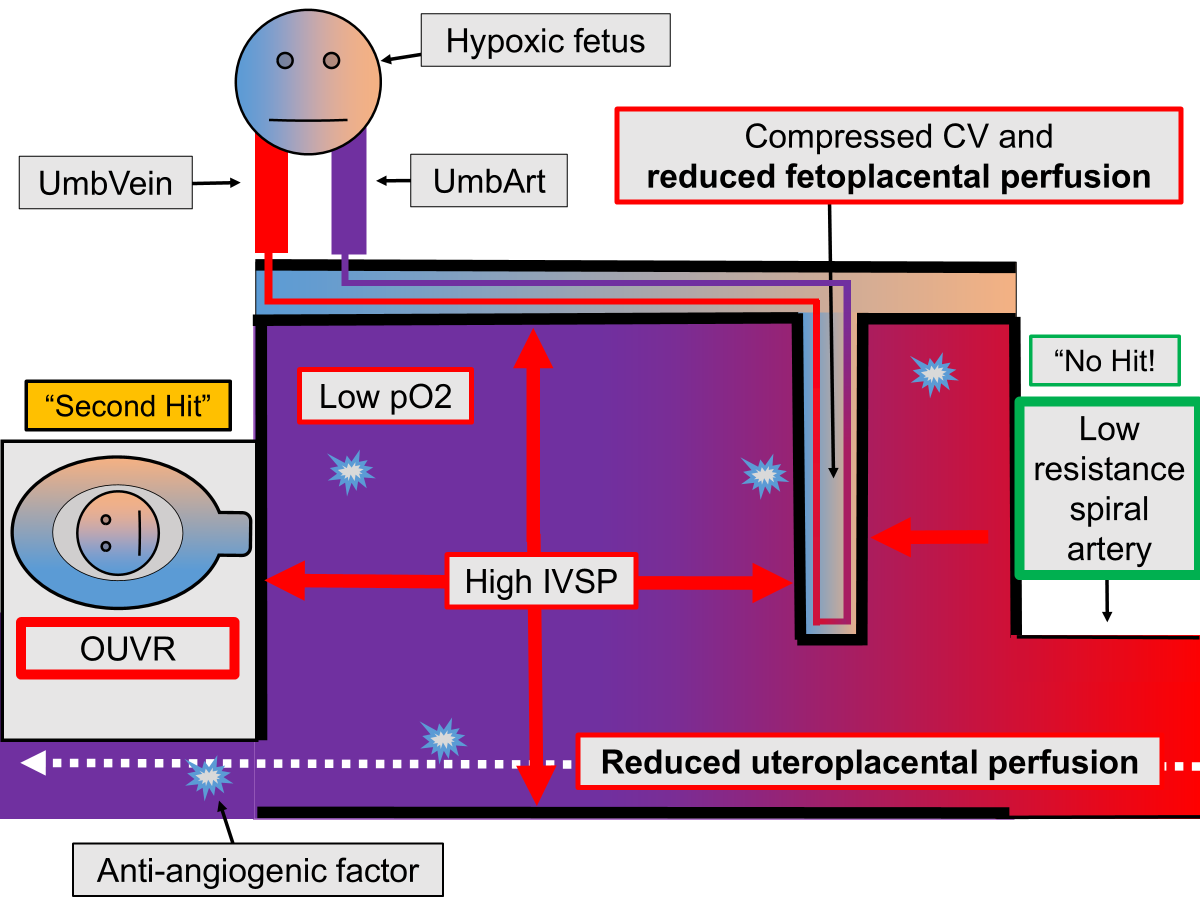
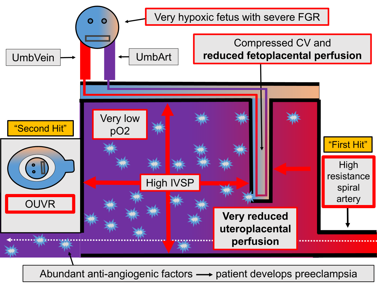
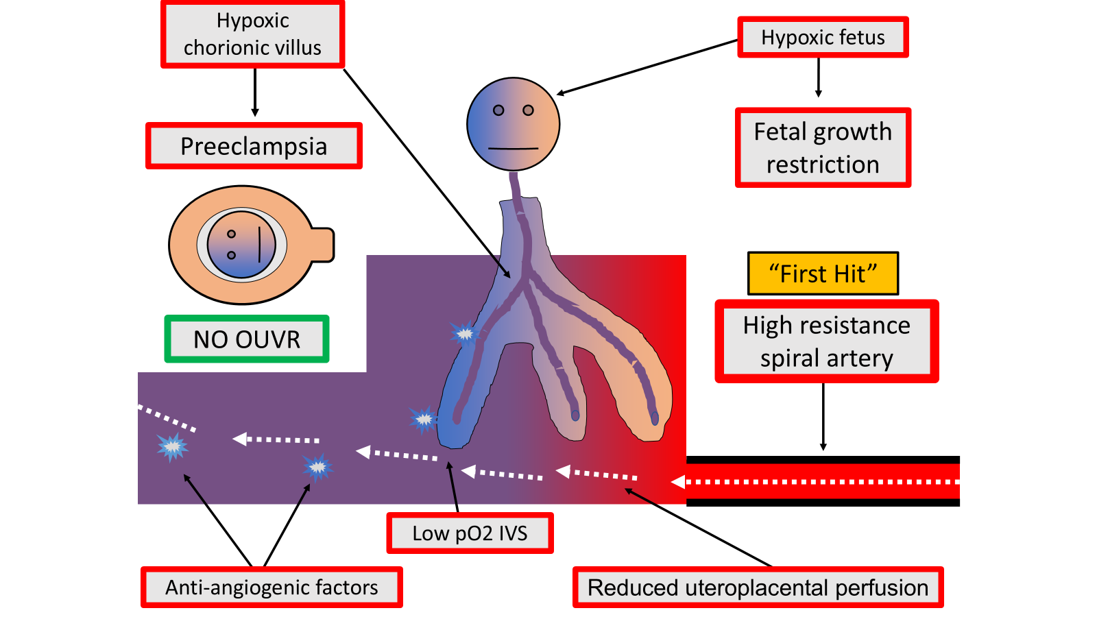
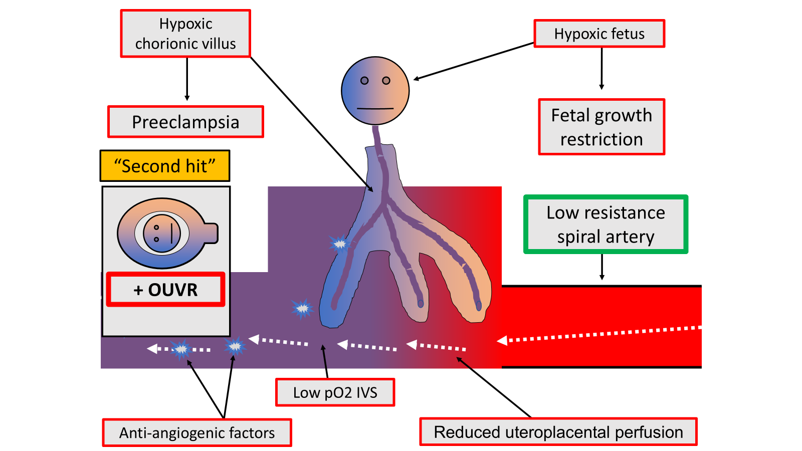
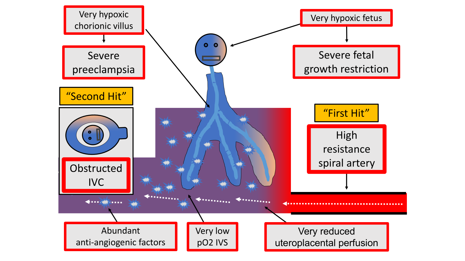
Anti-angiogenic factors — chemicals released into the mother’s bloodstream by the placenta when it is not getting enough oxygen. These chemicals are thought to cause preeclampsia, a dangerous complication of pregnancy, involving high blood pressure, body swelling and other serious organ damage in the mother—as well as possible fetal growth restriction (below normal growth of fetus).
Aorta — the main artery of the body, starting at the heart and carrying oxygen-containing blood to the entire body
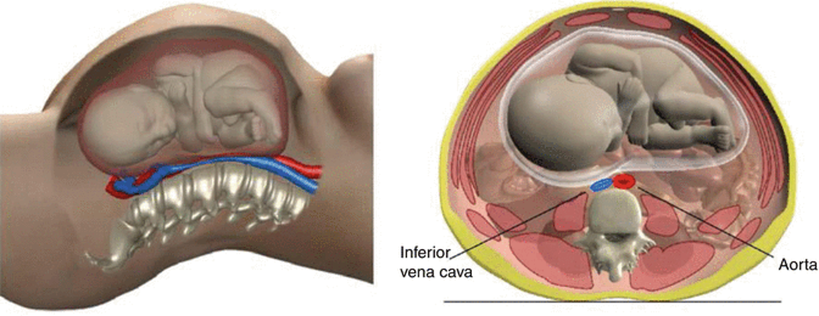
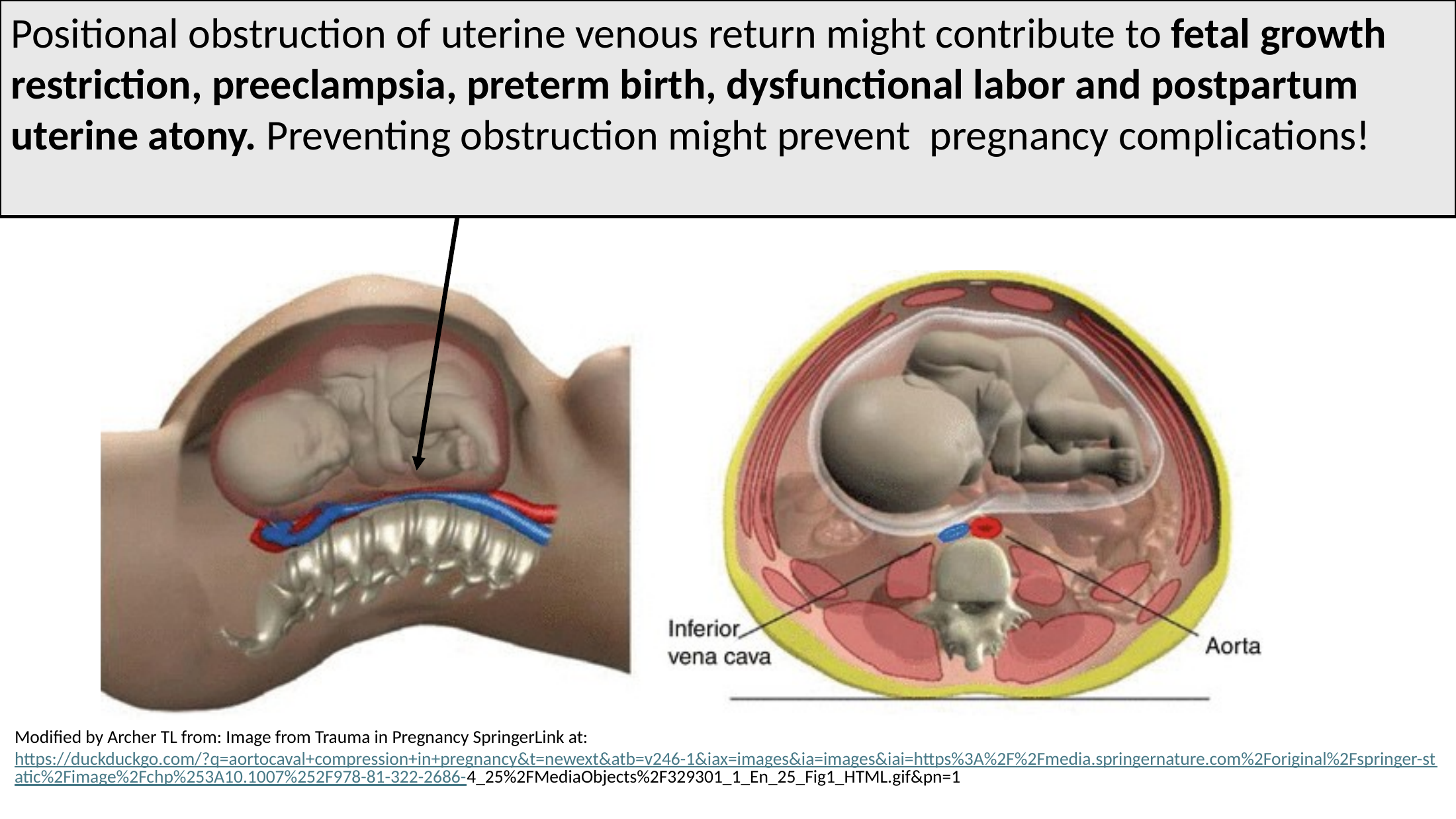
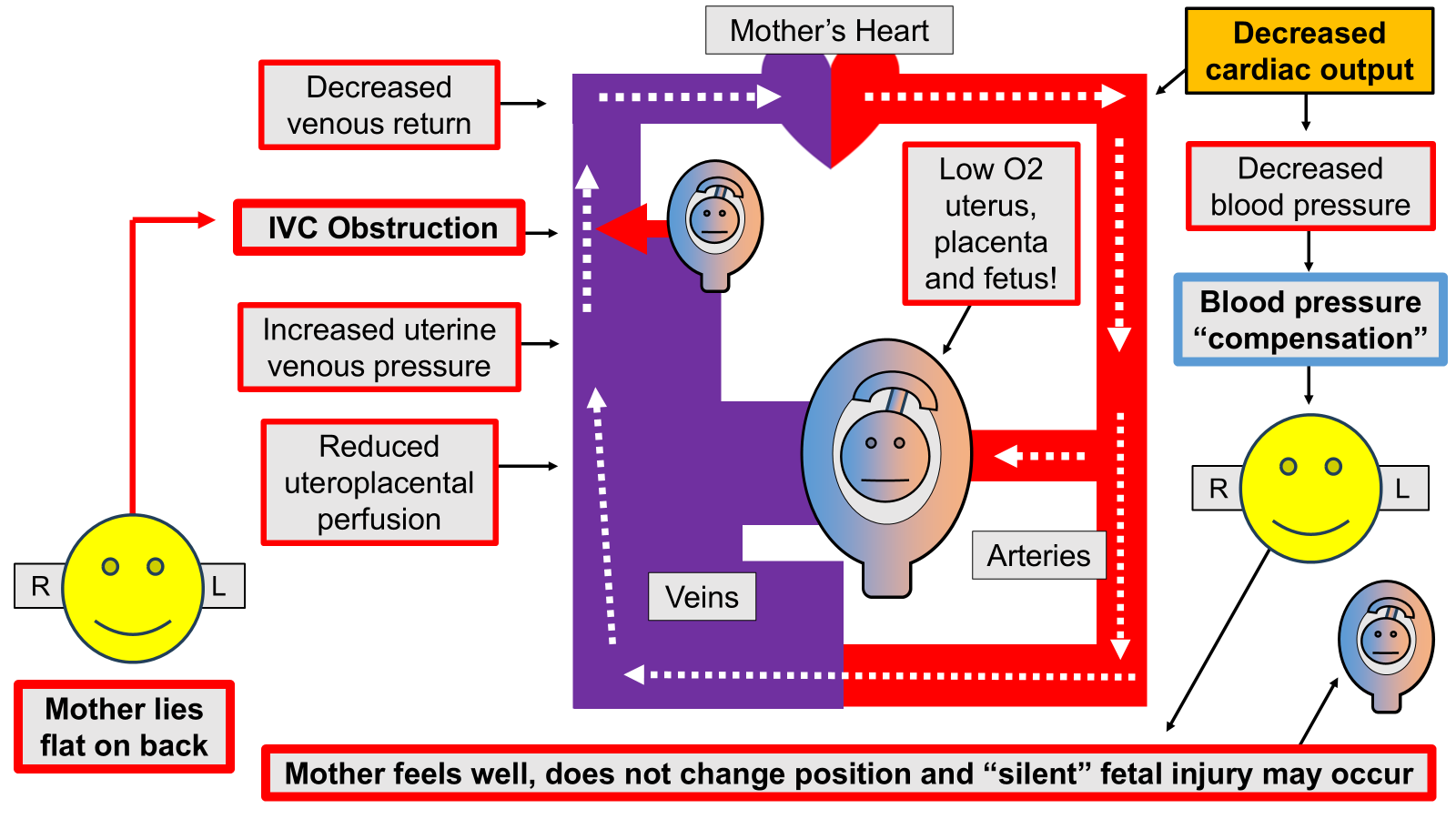
Aortocaval compression (ACC) — when the pregnant (“gravid”) uterus physically compresses and blocks blood flow through both the aorta (the main artery of the body, which carries oxygenated blood away from the heart to the organs of the body) and the inferior vena cava (the biggest vein in the abdomen, which carries blood back to the heart so it can pick up more oxygen in the lungs).
Arteries — the blood vessels taking well-oxygenated blood away from the heart and delivering it to the organs of the body
Autotransfusion — During labor, the uterus contracts to push the baby down the birth canal, and these contractions squeeze some of the mother’s blood which is inside the uterine muscle back into the mother’s active circulation. This autotransfusion causes a measurable increase in the mother’s cardiac output for a brief period of time. Autotransfusion can be detected and followed by using commercially available and non-invasive technology such as Electrical Cardiometry.
Blood pressure (BP) — the pressure (force per unit area) that the blood has while it is within an artery or vein. Usually, the term applies to the pressure of the blood within arteries, but the blood pressure within veins (the “venous pressure”) is also important within the body. Venous pressure that is too high can prevent organs from receiving blood flow and oxygen and can also cause organ swelling (called “edema”).
Cardiac output (CO) — The volume of blood the heart pumps out to the body each minute. The CO is measured in liters per minute and abbreviated as “L/min”
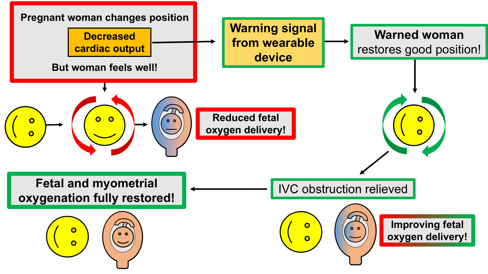
Cardiac output-guided maternal positioning (COGMP) — The use of positional decreases in the mother’s cardiac output to diagnose a mother’s body position which may be bad for the baby growing inside the mother (the “fetus”). The term is new and is not in wide use. This idea is one of the main new ideas of this website.
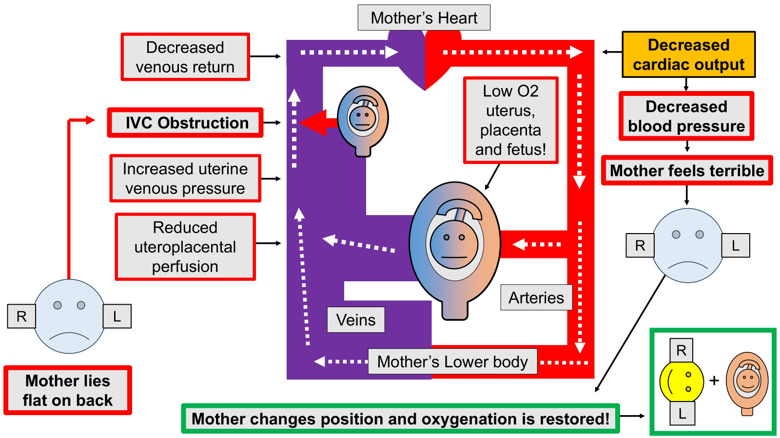
Cardiovascular compensation — The idea that the mother’s heart and blood vessel system (cardiovascular system) can keep the mother’s blood pressure in the normal range, even though the mother’s cardiac output has been decreased by obstruction of the main vein of the lower body, the inferior vena cava.
Cardiovascular system — The entire system of the heart, blood vessels and the nerves that control those organs. Regulation of the cardiovascular system is extremely complicated!
Cesarean delivery (CD) — The more modern term for “cesarean section,” or the delivery of the baby from the mother by means of surgery through the lower abdomen.
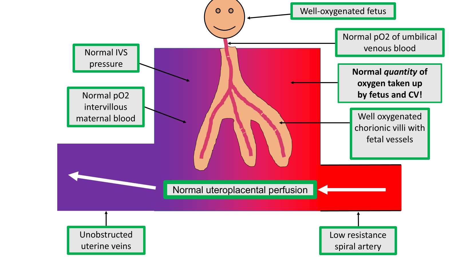
Chorionic villi / chorionic villus — The parts of the placenta (fetal tissue) that contain some of the fetus’ blood vessels, which pick up oxygen (by diffusion) from the mother. The bloodstreams of the baby and the mother normally do not mix to any significant extent, and all of the transfer of oxygen, carbon dioxide, food molecules and waste products to and from the fetus occurs by various types of transfer across tissue. The commonest form of transfer from one bloodstream to another is called “diffusion.”
Chronic — adjective describing something that happens over a long period of time. Used in contrast to “acute” which means something that occurs all of a sudden.
Doppler ultrasound — the medical use of sound waves to gather medical images and other types of medical information, such as the speed of blood flow in parts of the body.
Dysfunctional labor — When the contractions of the uterus during labor do not work well to push the baby down the birth canal. One common type of dysfunctional labor is when the uterine muscle (myometrium) loses strength during labor, and this type of dysfunctional labor can be caused by a lack of oxygen in the myometrium.
Electrical cardiometry (EC) — A type of electrical measurement of the cardiovascular system sold by Osypka Medical, Inc. Electrical cardiometry is one type or “brand” of what is called “impedance cardiography.” Other types of impedance cardiography are available commercially, but the type I used in my research was electrical cardiometry.
Electrical velocimetry — A term closely related to “electrical cardiometry” which means “measuring the speed of blood flow.”
Endometritis — Infection of the inside of the uterus occurring after delivery of the baby
Epidural analgesia (“an epidural”) — A type of pain relief administered to a pregnant woman to relieve labor pain. It is given into a mother’s back through a tiny, flexible tube.
“Fetal distress” — An older term that has largely been replaced by the term “non-reassuring fetal heart tones.” The condition is usually diagnosed by a slowing of the fetus’ heart rate, and this slowing is sometimes caused by a lack of oxygen in the fetus.
Fetal growth restriction (FGR) — An abnormally low amount of fetal size or growth. The term used to be called “fetal growth retardation,” but was changed because the word “retardation” suggested the phrase “mental retardation.”
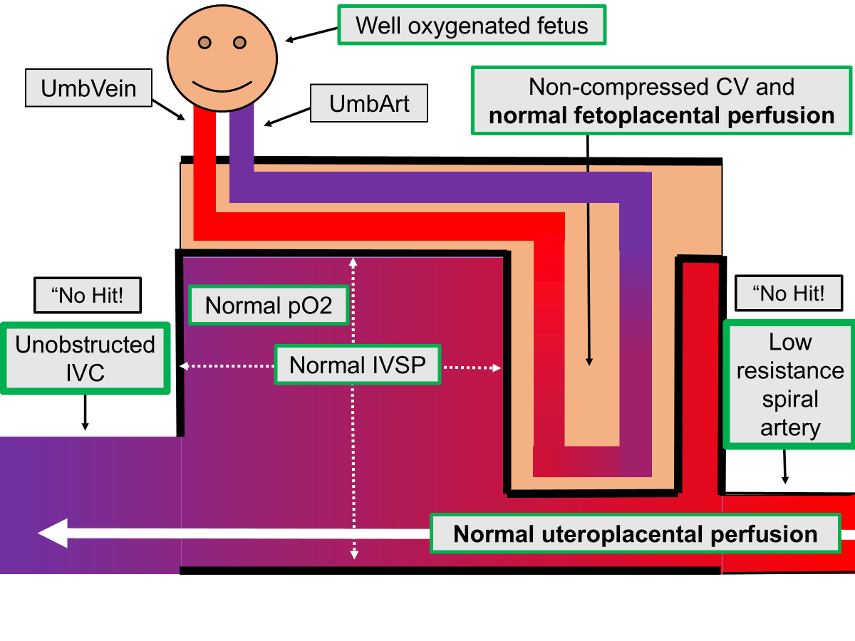
Fetoplacental perfusion — the blood flow to the placenta coming from the fetal heart. This blood flow is completely separate from the “uteroplacental perfusion” which is blood flow coming from the mother’s heart. Exchange of gases, food molecules and waste products occurs from one bloodstream to another across the tissue of the chorionic villi (the “placental barrier”) by diffusion and other mechanisms.
Fetus / fetal — The term for the baby while it is developing inside the mother’s uterus, before delivery. Once the fetus comes out of the mother, the fetus is called a “baby.”
Gravid uterus — “Gravid” means “pregnant” or “carrying a fetus.” So, “gravid uterus” means a woman’s uterus with a developing fetus inside. When the fetus becomes large during the second half of pregnancy, the gravid uterus can block blood flow back to the mother’s heart through the major vein of the lower body (the “inferior vena cava”)
Hemodynamics / hemodynamic — means the study or process of how the heart pumps oxygen-carrying blood to the organs of the body and back again to the heart, for re-oxygenation in the lungs. Another term for the flow of blood within the body.
Hemorrhage — Uncontrolled or unwanted loss of blood from blood vessels
Hypotensive / hypotension — means “having a blood pressure which may be too low for health”
Hypoxemia — low and unhealthy amounts of oxygen in blood.
Hypoxia / hypoxic — refers to an unhealthy low level of oxygen in a tissue organ or individual
Inferior vena cava (IVC) — the main large vein carrying venous blood from the lower body back to the heart. The pregnant (“gravid”) uterus can block the IVC within the abdomen during the second half of pregnancy.
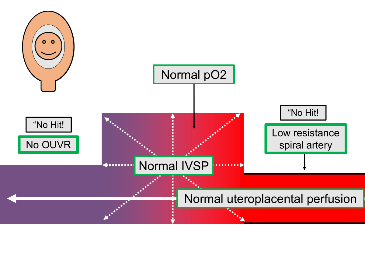
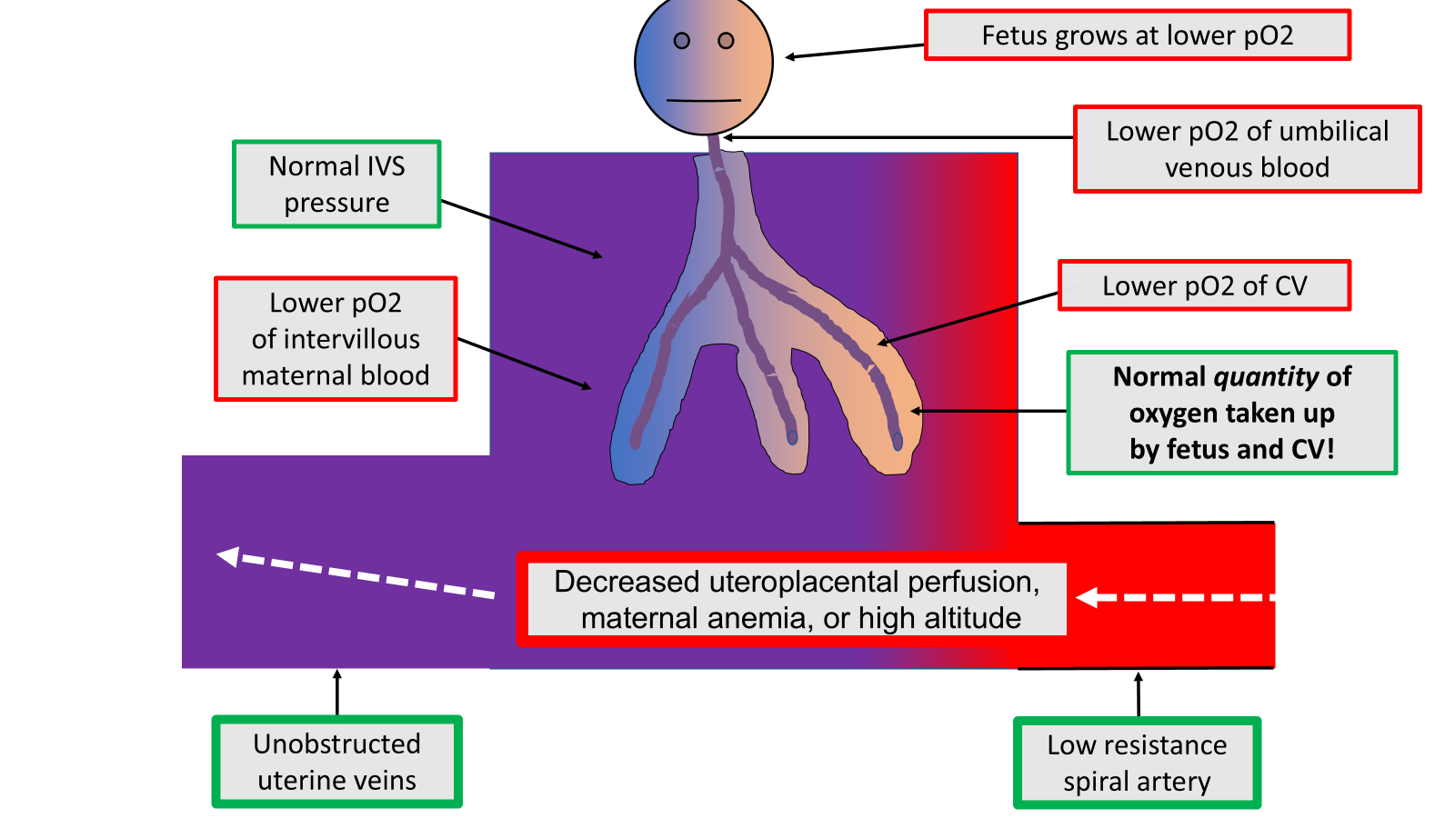
Intervillous blood flow — the flow of maternal blood around the chorionic villi within the placenta. The blood enters the “intervillous space” through the “spiral arteries” and leaves the space through the uterine venous system, which can be blocked by the pregnant uterus.
Intervillous perfusion — another name for “intervillous blood flow,” the flow of maternal blood around the chorionic villi within the placenta. “Perfusion” means the flow into and then out of an organ.
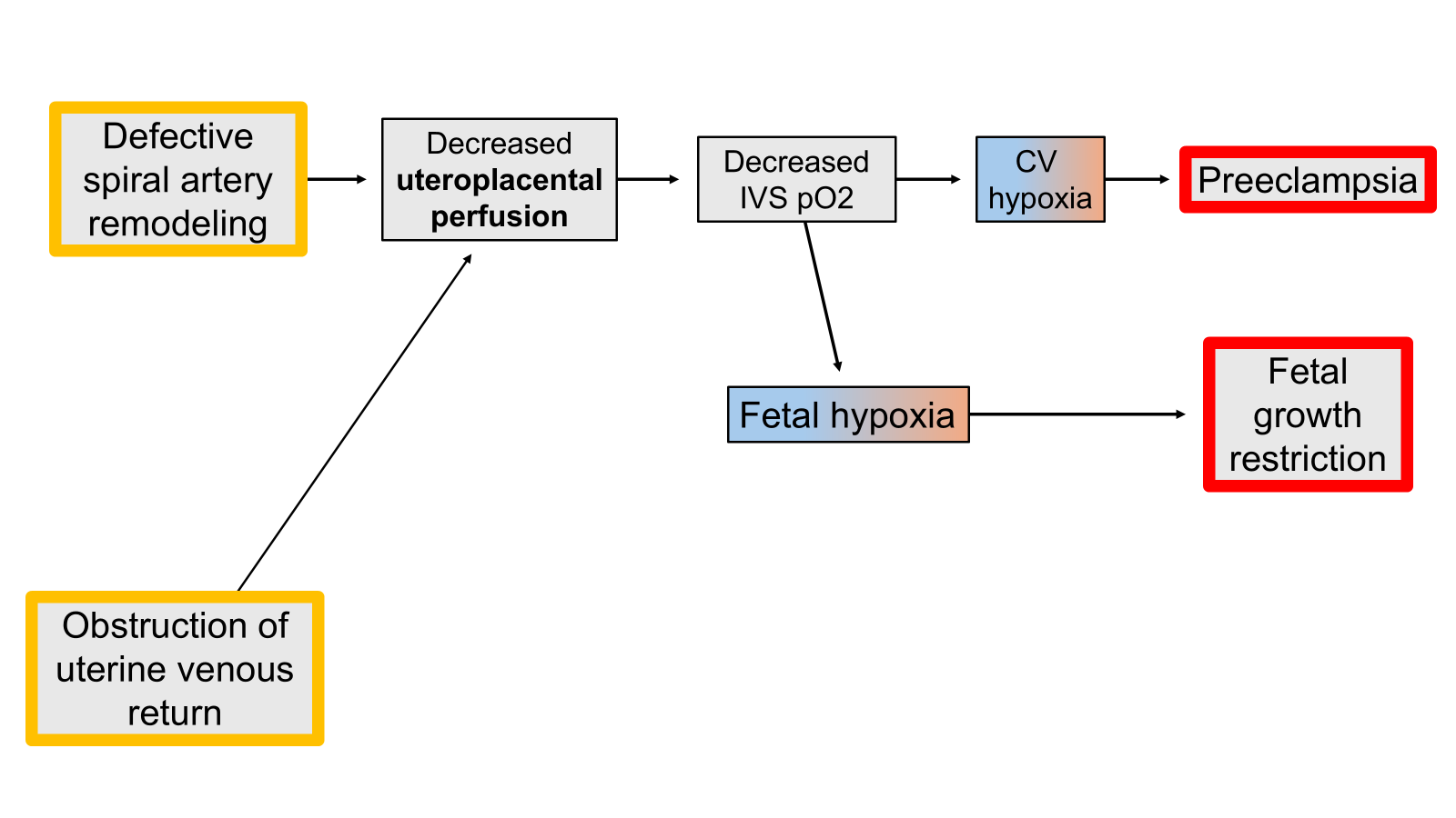
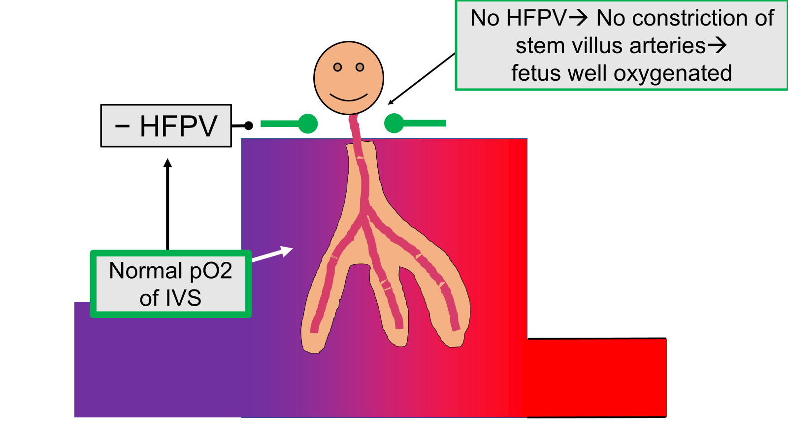
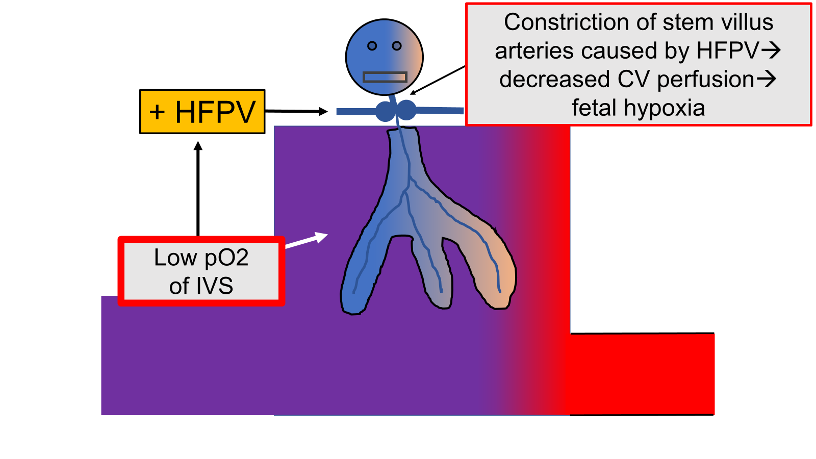
Intervillous space (IVS) — The space or volume containing the maternal blood flows around and surrounds the chorionic villi, which are the “fingers” of fetal placental tissue containing the fetal blood vessels. The flow of blood (“perfusion”) through the IVS starts with blood coming out of the maternal “spiral arteries” and ends when that blood enters the maternal “spiral veins” which are the start of the uterine venous return system which carries blood back to the mother’s heart.
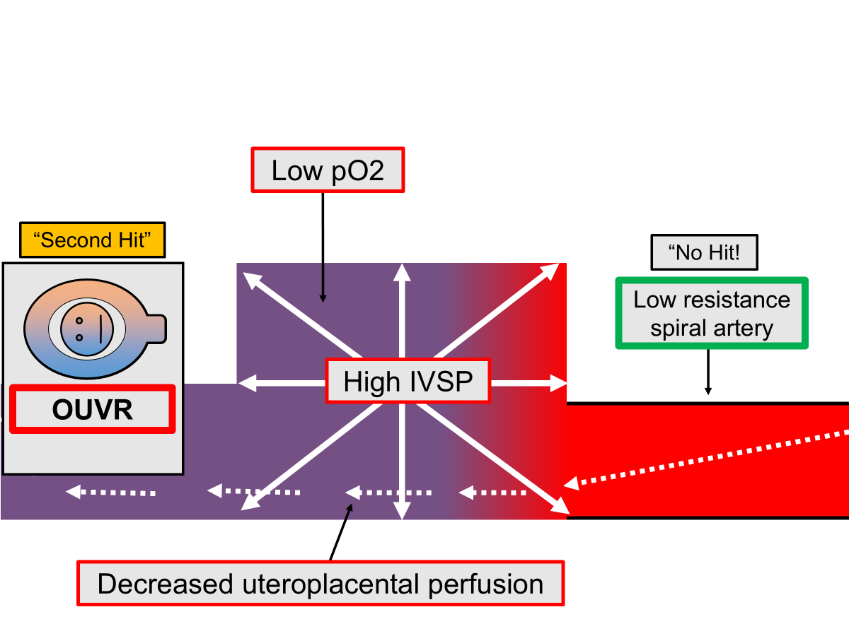
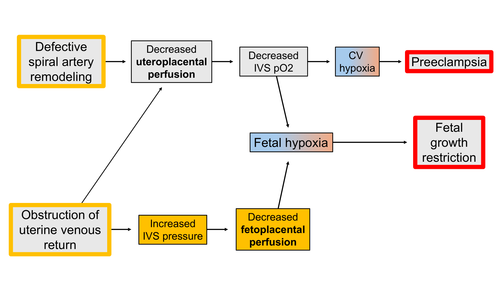
Intervillous space pressure (IVSP) — The pressure of the blood within the intervillous space. When the intervillous space pressure increases due to obstruction of the uterine venous system, it can compress the chorionic villi and reduce fetoplacental perfusion—which could be dangerous for the fetus.
Ischemia / ischemic — describes the situation when an organ is not getting all the blood flow (“perfusion”) it needs to stay healthy.
IVC obstruction — obstruction (blockage) of the inferior vena cava
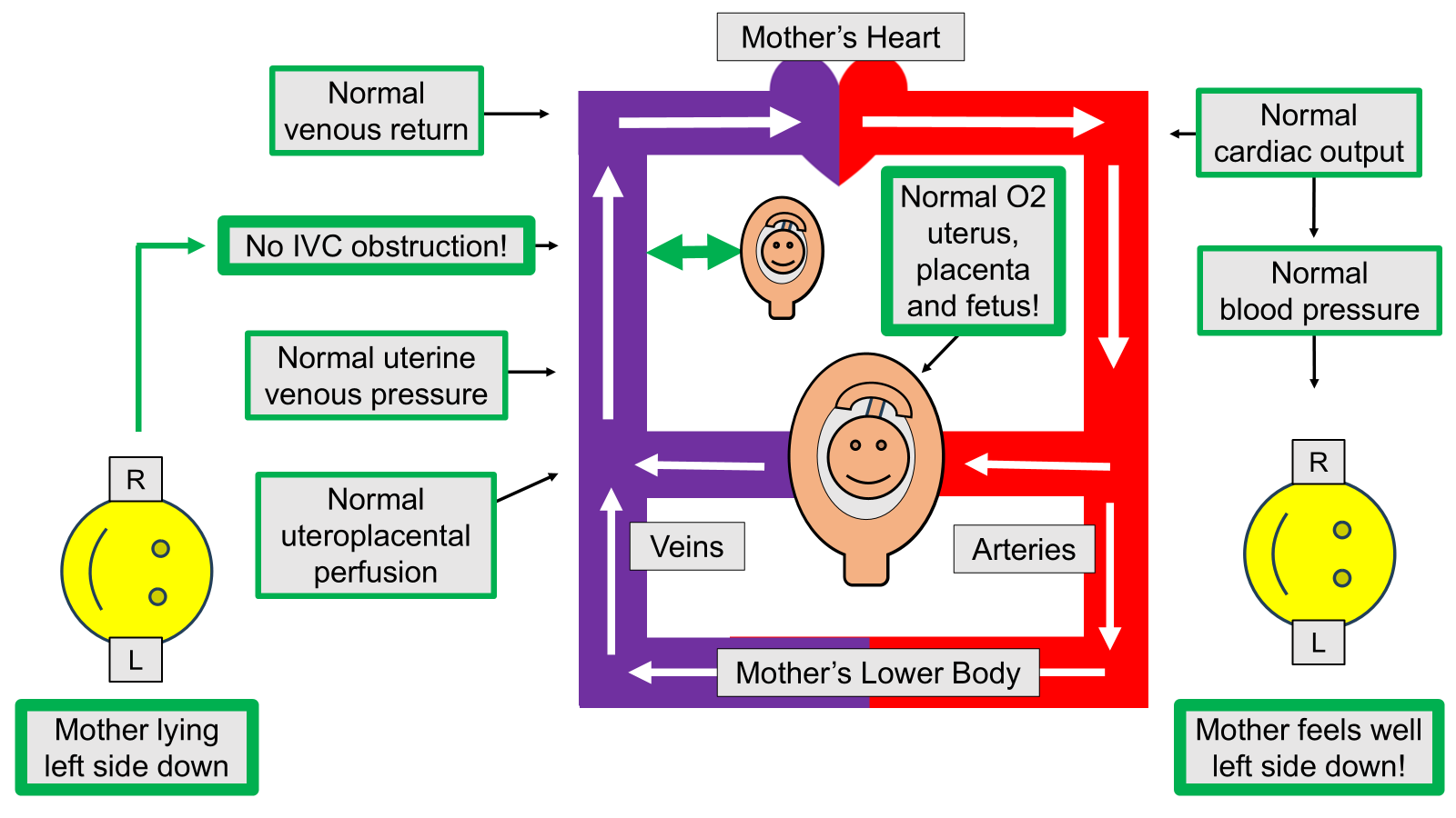
Lateral position — when the mother lies on one side or the other. Left lateral means the left side of the body is down, whereas right lateral means that the right side of the body is down.
Maternal — refers to the pregnant woman who is going to give birth to a baby
Maternal–fetal medicine (MFM) — that sub-specialization within the specialty of Obstetrics and Gynecology which refers to the advanced care of the pregnant woman and fetus, considering both mother and fetus as “patients” and as a closely-coupled pair of individuals. MFM specialists are trained obstetrician-gynecologists (OBGYNs) who then receive several more years of advanced training after becoming OBGYNs in order to become MFM specialists—who are often called “perinatologists.”
Myometrial ischemia — lack of adequate blood flow and oxygen delivery to the smooth muscle of the uterus which contracts during labor. Myometrial ischemia can be a cause of poor uterine contractions during labor.
Myometrium / myometrial — Refers to the smooth muscle of the uterus which normally contracts strongly during normal labor.
Non-invasive — refers to medical monitoring or measuring techniques or devices which do not require any insertion of anything into the body across the skin, such as small plastic tubes or “cannulae.” Non-invasive techniques are usually pain-free, easier to perform and much less risky than “invasive” techniques such as sticking plastic tubes into blood vessels through the skin.
Normoxic — means that the amount of oxygen in something (e.g. blood or tissue) is at a normal and healthy level
Obstetric anesthesia — a subspecialty of Anesthesiology and Critical Care, which has to do with the care of pregnant women and their fetuses.
Obstetrician — a doctor who takes care of pregnant women
Obstetrics / obstetric — the medical care of pregnant women
Obstruction of uterine venous return (OUVR) — a key phrase for this website, meaning any blockage to the de-oxygenated venous blood coming out of the uterus that is “trying” to get back to the heart. A key point of this website is that OUVR can increase pressure within the placenta which can reduce oxygen delivery to the developing fetus.
Oxygen (O2) — the gas in our air that is needed for life by all animals. Air contains 21% oxygen, with almost all the rest being nitrogen (N2).
Oxygen partial pressure (pO2) — the amount or pressure of oxygen within a gas or a liquid such as blood.
Oxygenation — the process of putting oxygen into a tissue to enable it to live. Normally, tissues are “oxygenated” when they receive the flow of oxygen-carrying blood.
Oxytocin — a medication used in obstetrics to increase the frequency or strength of uterine contractions, usually during labor. Oxytocin also occurs naturally in the body, and is released when the baby sucks on the mother’s nipple during breastfeeding.
Perfusion — the successful flow of blood into and out of an organ. Perfusion normally causes oxygenation of tissue.
Perfusion pressure — the mathematical difference between the arterial pressure and the venous pressure for an organ. An increase in venous pressure with a constant arterial pressure decreases the perfusion pressure.
Perinatology — the field of medicine concerned with the advanced medical care of pregnant women and their fetuses. Perinatologists receive further years of advanced training after becoming obstetrician-gynecologists (OBGYNs). The field of perinatology is also called “maternal fetal medicine” or MFM.
Placenta / placental — the placenta is almost entirely fetal tissue, with just a tiny amount of maternal tissue (the “decidua”) breaking away from the uterus at the time of delivery. The placenta is the organ that allows exchange of everything (oxygen, carbon dioxide, food molecules, waste molecules, signaling molecules, etc.) between the pregnant woman and her fetus during pregnancy. The separate circulations of the fetus and the mother do not mix during normal pregnancy (with rare exceptions), and all of the exchange between fetus and mother occurs across the part of the chorionic villi which is called the placental membrane.
Positional fetal oxygenation management — the term (which I believe I invented) to describe the use of maternal positioning to enhance the delivery of oxygen to the fetus.
Postpartum — after delivery
Postpartum hemorrhage — excessive maternal bleeding after delivery of the baby
Postpartum uterine atony — inadequate contraction of the uterus after delivery. This website presents the theory that poor myometrial oxygenation during labor can be a cause of postpartum uterine atony.
Preeclampsia — A dangerous syndrome occurring after 20 weeks of pregnancy, involving maternal high blood pressure, body swelling and maternal organ injury and dysfunction, such as having protein in the urine and decreased kidney function. Preeclampsia has been the subject of intensive study for more than a century, and much progress has been made in its diagnosis and treatment, but it is still poorly understood and a major pregnancy problem whose incidence is not decreasing.
Prenatal care — the process of care of the pregnant woman before birth
Preterm birth — birth before 37 completed weeks of pregnancy
Preterm labor — onset of labor before 37 completed weeks of pregnancy
Spiral arteries — the small maternal arteries that eject maternal blood into the intervillous spaces of the placenta, so that the blood can come in contact with the chorionic villi and give oxygen to the fetus. A major theory of preeclampsia is that it has to do with “inadequate remodeling” of the spiral arteries, resulting in an inadequate release of maternal blood into the intervillous spaces.
Spiral artery remodeling — the process by which the spiral arteries (see above) are converted from high resistance arteries to low resistance arteries, which increases the flow rate of maternal blood into the intervillous spaces to satisfy the fetus’ growing need for oxygen.
Supine hypotensive syndrome of pregnancy — the maternal syndrome of pallor, dizziness, nausea and vomiting, rapid heart rate, low blood pressure and possible loss of consciousness that sometimes happens to pregnant women late in pregnancy who lie flat on their backs (supine). The syndrome is caused by the uterus blocking a lot of blood from getting back to the heart and this causes a serious decrease in blood pressure, which then causes the syndrome.
Systemic vascular resistance — the total resistance of (mostly) the arterial system to the blood pumped out by the heart. You can think of it like putting your thumb over the mouth of a garden hose: the partial blockage of flow increases the pressure of the stream of water, but decreases the amount of water that actually comes out of the hose.
Transthoracic echocardiography — The use of high-frequency (inaudible) sound waves to form moving images of the heart. Over the last 50 years, it has become a key medical procedure for “looking at” the beating heart in real time.
Two-hit hypothesis — the idea that it takes two problems to cause a major problem. In other words, that two different problems cause much more damage together than either problem alone. In this website I present the hypothesis that obstruction of uterine venous return (OUVR) can act as a “second hit” and make other threats to fetal oxygenation (such as poor spiral artery remodeling) much more dangerous. This example may be important because OUVR can be prevented-- even though the “first hit” may not be preventable at all.
USCOM (Ultrasound cardiac output monitor) — a commercially available device which uses sound waves to “look at” the blood flow within the ascending aorta to see how fast the blood is flowing out of the heart and how long the flow lasts. The device “looks at” the blood flow in the ascending aorta through an “ultrasound window” that lets sound waves pass through it which is located at the top of the breastbone at the base of the neck. The “ultrasound window” is called the “sternal notch” and you can feel it on yourself with your finger.
Ultrasound — high frequency sound waves used for “looking” inside the body and creating images of structures like the heart, liver or blood vessels.
Umbilical artery — the fetal artery which carries less oxygenated blood from the fetus to the placenta, where the blood picks up more oxygen to meet the needs of the fetus.
Umbilical vein — the fetal vein which brings more oxygenated blood back to the fetus from the placenta
Uterine atony — poor tone (or degree of constant contraction) of the uterus after delivery. Uterine atony often causes postpartum hemorrhage, and this website presents the theory that postpartum uterine atony could be caused—at least in part—by poor oxygenation of the myometrium during labor, due to obstruction of uterine venous return (OUVR) during labor.
Uteroplacental perfusion — the blood flow to the placenta coming from the mother’s heart. This blood flow is completely separate from the “fetoplacental perfusion” which is blood flow coming from the fetus’ heart. Exchange of gases, food molecules and waste products occurs from one bloodstream to another across the tissue of the chorionic villi (the “placental barrier”) by diffusion and other mechanisms.
Veins — the thin-walled blood vessels that normally carry de-oxygenated blood back to the heart, so that the blood can be pumped to the lungs by the right ventricle to pick up more oxygen.
Venous pressure — normally, venous pressure is very low compared to arterial pressure, and the difference between the two pressures across an organ is called that organ’s “perfusion pressure.” When venous pressure increases, it causes two potential problems: 1) decreased organ perfusion, and 2) swelling of the organ, with edema formation and resulting organ dysfunction.
Venous return — a global term used to describe the total amount of venous blood that comes back to the heart, from both inferior and the superior venae cavae. A very important insight is that—in a steady state when everything is stable—the cardiac output is the same as the venous return. After all, the heart can only pump out all the blood that it receives from the veins! A corollary of this fact is that when the uterus blocks the inferior vena cava within the abdomen it blocks the venous return and the maternal cardiac output decreases. These positional decreases of maternal cardiac output can be used to detect and correct the obstruction of uterine venous return that occurs when the inferior vena cava is obstructed.