Could your body position affect oxygen delivery to your unborn baby?
This article presents the theory that unfavorable maternal body positions during the second half of pregnancy might deprive the developing fetus of all the oxygen required for optimal fetal development, and that this periodic oxygen deprivation might cause long-lasting fetal and adult injury. This same oxygen deprivation of the fetus, placenta and myometrium-- caused by obstruction of uterine venous return by the gravid uterus itself in certain maternal positions-- might also contribute to the causation of preeclampsia, fetal growth restriction, preterm birth, dysfunctional labor, cesarean delivery, fetal distress during labor and postpartum uterine atony and hemorrhage. The purpose of this article is to stimulate further research and discussion, since prevention of the problem-- if it is shown by future research to be a significant source of injury-- might represent “low-hanging fruit” in efforts to improve pregnancy outcomes for both mothers and their babies. This article presents an unproven theory and is not meant to alarm anyone, but I believe that there does exist sufficient evidence to suggest that this idea may have merit and therefore that the idea deserves more research attention than it is currently receiving. For individual pregnancy concerns, consult your obstetric care team.
Everyone acknowledges the importance of prenatal care, and it is widely accepted that pregnancy involves two “patients”—the mother and the fetus. For the benefit of both, pregnant women are urged to take their vitamins and iron, eat a healthy diet, get exercise and watch their weight. They will also be checked for anemia, high blood pressure, diabetes and urinary tract infections. Even more tests may be performed to detect Down syndrome or other genetic and anatomical abnormalities, and most people are aware of the dangers to the fetus of smoking, alcohol and many other drugs and legal medications.
But in our attempts to optimize fetal growth, development and lifelong health for both baby and mother, could it be that we are overlooking a threat to an absolutely essential “input” for the “production” of a healthy person— namely, an adequate supply of oxygen throughout pregnancy?
The fetus gets all of its oxygen from the mother via the placenta, as represented schematically in Figure 1. This Figure shows the ideal situation for fetal, placental and uterine oxygenation during the second half of pregnancy, when the uterus is large enough to obstruct crucial portions of the mother's venous system, but does not do so. Figure 1 shows maternal blood flow in a favorable maternal body position—by which I mean, a body position in which the heavily pregnant uterus is not compressing major maternal pelvic and abdominal veins.
But Figure 2 shows what happens when the mother changes her body position and the pregnant uterus does compress the major veins that carry blood back to the heart from the uterus and the entire lower half of the mother’s body. In this situation (labeled “IVC obstruction”) two bad things happen: 1) within minutes the amount of blood returning to the mother’s heart decreases, along with her blood pressure, and 2) the pressure increases within the veins draining the uterus, and this increased venous pressure decreases total blood flow and oxygen delivery to the uterus, placenta and fetus.
The situation shown in Figure 2 was first described in 1953 as the “supine hypotensive syndrome of pregnancy” and consists of maternal distress, pallor, sweating, low blood pressure, nausea, vomiting and an increase in venous pressure in the veins in the groin, when susceptible patients late in pregnancy lie flat on their backs (1). The original description of the syndrome did not emphasize the dangers inherent in the syndrome to the fetus, but rather emphasized the fact that promptly turning the patient onto one side relieved the maternal effects of the syndrome. Thus, the original conception of the syndrome was that of a short-term threat to the mother which could be easily corrected by turning the mother onto her side and thereby relieving the venous obstruction caused by the pregnant uterus.
A corollary of this perspective, however, is that the syndrome forces the mother or her care givers to change her body position, which then restores uterine and placental blood flow and fetal oxygenation—as well as blood flow to the mother’s brain. Hence, the syndrome —although highly unpleasant and dangerous for the mother— can also be seen as a protective response which protects both mother and fetus.
But what happens if the mother’s cardiovascular system compensates for the unfavorable situation shown in Figure 2 by increasing the mother’s blood pressure despite the reduction in cardiac output? This situation-- which is thought to occur commonly-- is shown in Figure 3, in which the mother continues to feel well because her blood pressure is maintained, but the blood flow and oxygen delivery to the uterus, placenta and fetus are still impaired. If this situation occurs, the mother’s body position continues to impair oxygen delivery to her developing baby without the mother being aware that anything is wrong! This prolonged deprivation of fetal, placental and uterine oxygenation might cause permanent injury.
Since 2011 I have authored many medical articles and two books, and spoken at four maternal-fetal medicine conferences in the UK, Italy and Slovenia. In these venues I have urged medical researchers to look further into this possible cause of unrecognized (or “silent”) fetal, placental and uterine oxygen deficiency during the second half of pregnancy. I have also argued that this occult injury might be a contributing factor to preeclampsia, fetal growth restriction, preterm birth, dysfunctional labor, cesarean delivery and postpartum hemorrhage (2-17). Many well-recognized maternal-fetal medicine specialists have shown what appears to be genuine interest in my ideas, but so far no further research has been carried out to specifically evaluate my hypotheses. In 2015 I retired from the University of California San Diego as Clinical Professor of Anesthesiology and Director of Obstetric Anesthesia, so I can no longer initiate any more clinical studies on my own, although I have offered to help other investigators with new research. This lack of follow-up has been particularly frustrating since I have also proposed a possible method of preventing possible silent fetal and maternal injury due to a lack of oxygen in the uterus, placenta and fetus. I have called this proposed technique “cardiac output-guided maternal positioning” since it would use the tell-tale reduction in maternal cardiac output as an actionable warning signal for the mother to change her body position in real time to restore unobstructed blood flow and oxygen delivery to her reproductive system. This use of positional decreases in maternal cardiac output as a warning signal to protect her baby from a “silent threat” is shown in Figure 4, and this preventive warning system might eventually be embodied in wearable medical devices such as “bracelets,” “rings” or “earrings” which would continuously and non-invasively monitor the pregnant woman’s cardiovascular system and give the mother a discreet warning when her body position appeared to be causing a possible problem for the fetus.
This article presents a theory which needs additional research to either demonstrate or disprove its validity and value. But if this theory is true, the preventive approach it suggests might improve the lifelong health of both mothers and their children, by ensuring that developing babies will always get all the oxygen they need in order to develop to their full potential.
Figure 1Ideal maternal blood flow to and from the pregnant uterus and lower body
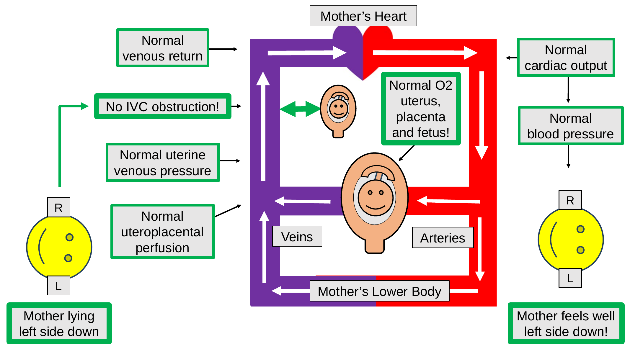
Ideal maternal blood flow to and from the pregnant uterus and lower body during the second half of gestation when the uterus is large enough to obstruct major maternal pelvic and abdominal veins, but is not doing so, because the mother has adopted a favorable body position which prevents major venous obstruction by the mobile uterus.
Abbreviations: IVC inferior vena cava, O2 oxygen
Figure 2The supine hypotensive syndrome of pregnancy
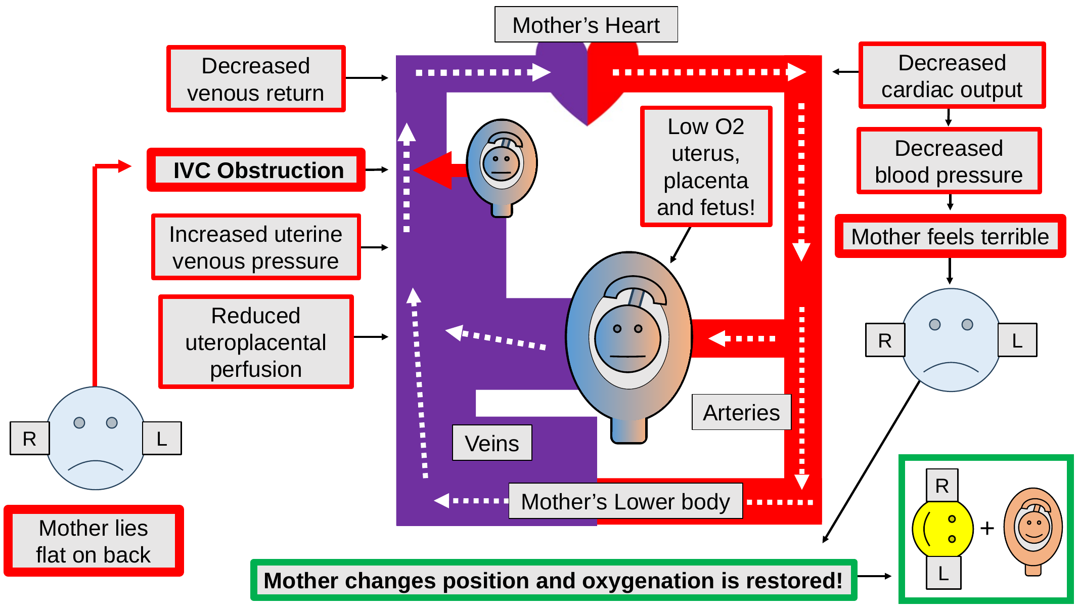
The “supine hypotensive syndrome of pregnancy” represents a threat to the oxygenation of both the mother and the fetus, but it is usually regarded as just a short-term maternal problem which can be corrected by a prompt maternal position change from supine (flat on the back) to a lateral position. But during the syndrome, the fetus is in trouble as well as the mother, and changing the mother’s position may save both the mother and the fetus from injury due to a lack of oxygen. The green-bordered text box in the lower right-hand corner of the Figure 2 shows the condition existing after the beneficial maternal position change has been performed.
Abbreviations: IVC inferior vena cava, L patient’s left side, O2 oxygen, R patient’s right side
Figure 3Maternal cardiovascular compensation for reduced cardiac output
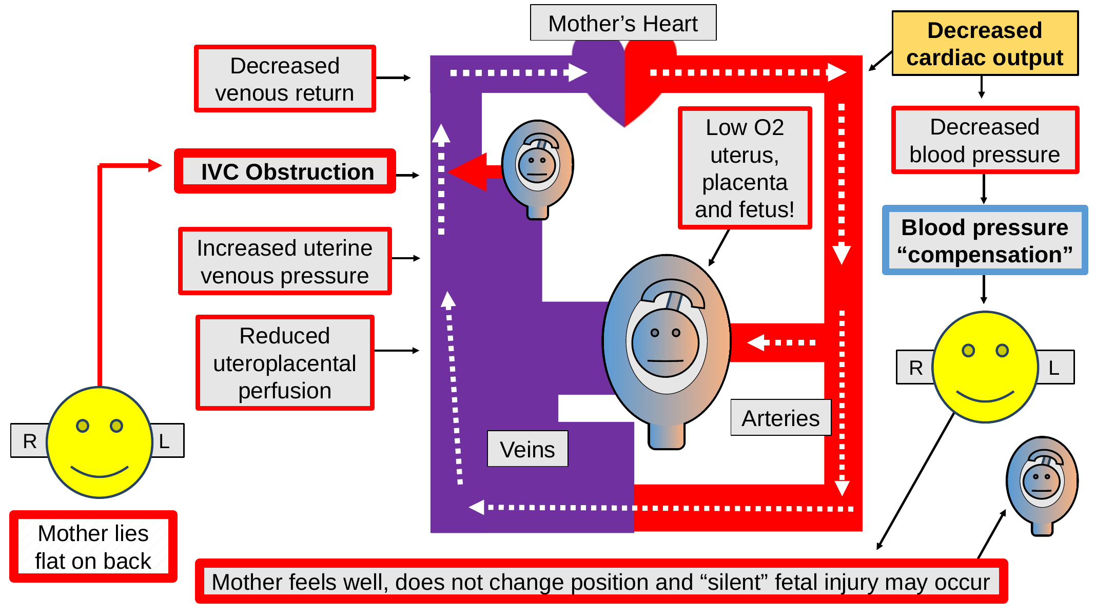
Maternal cardiovascular compensation for reduced cardiac output, with resulting normal maternal blood pressure and a maintained maternal sense of well-being. But this “compensation” would be a “false friend” since it would deceive the mother into thinking that all was well with her pregnancy. In this Figure we see that although the mother feels well, her fetus, placenta and uterus may not be getting all the oxygen they need for optimal development and function. The blood flow condition shown might contribute to preeclampsia, fetal growth restriction, preterm birth, dysfunctional labor, cesarean delivery and postpartum hemorrhage, but this is just a theory, and more research is needed to evaluate this hypothesis.
Abbreviations: IVC inferior vena cava, L patient’s left side, O2 oxygen, R patient’s right side
Figure 4Cardiac output-guided maternal positioning
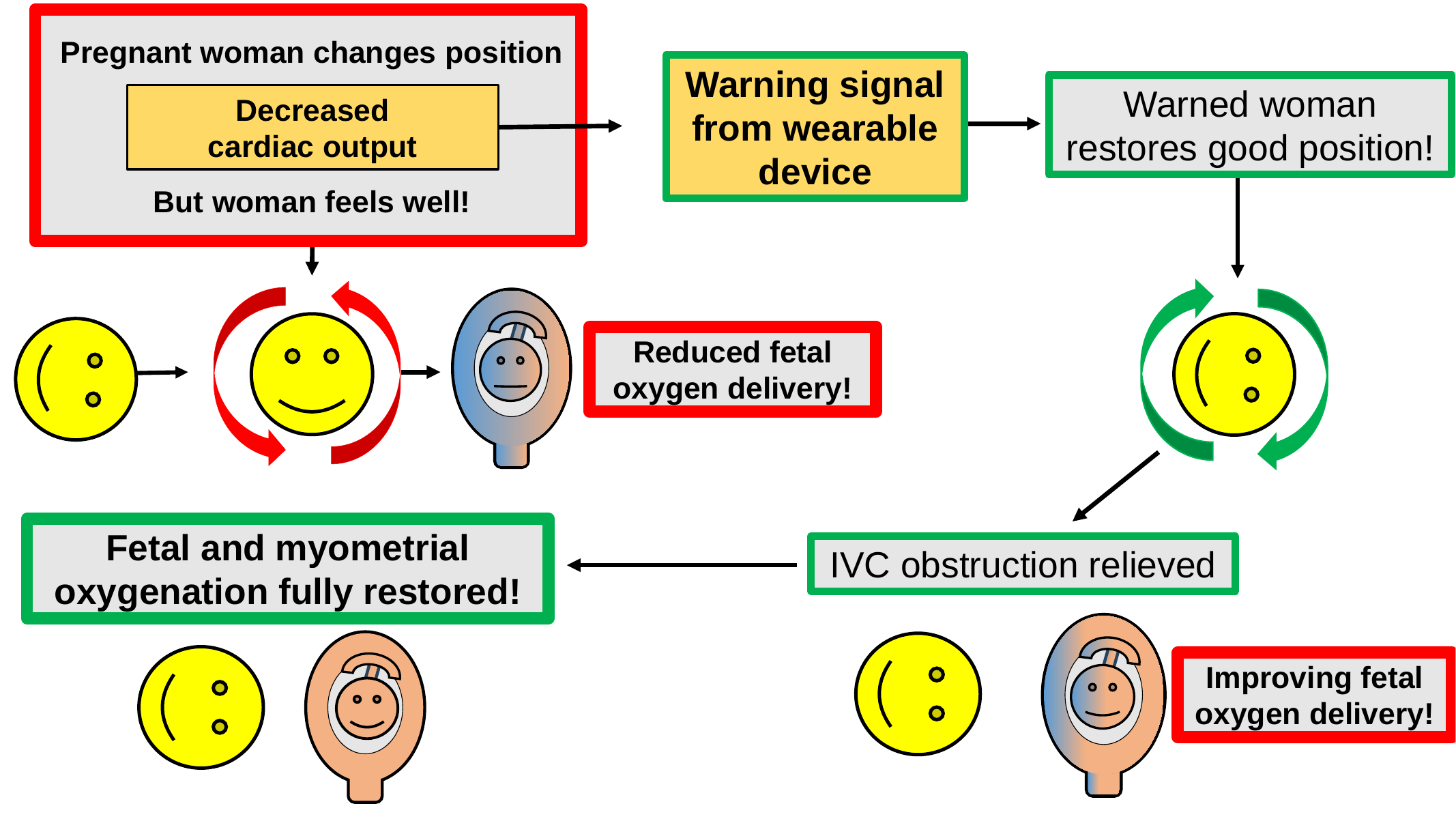
A positional decrease in maternal cardiac output may be able to serve as an actionable warning signal to the mother to change her body position to restore unobstructed blood flow and full oxygenation to her fetus and the rest of her reproductive system, even though she may feel comfortable in her current body position. I have named this concept “cardiac output-guided maternal positioning.” The positional decreases in maternal cardiac output (which are known to occur) may be detectable using wearable and completely non-invasive sensors in the form of “rings,” “bracelets” or “earrings.” This Figure is speculative and much further research is needed to confirm or disprove the concepts shown.
Abbreviations: CO cardiac output, IVC inferior vena cava
References
1. HOWARD BK, GOODSON JH, MENGERT WF. Supine hypotensive syndrome in late pregnancy. Obstet Gynecol. 1953 Apr;1(4):371-7. PMID: 13055188.
2. Archer TL. Therapeutic trials of maternal position therapy are needed to clarify the causes of abnormal maternal hemodynamic profiles and reduced umbilical vein flow in fetal growth restriction. J Perinat Med. 2026 Jan 14;54(2):431-432. doi: 10.1515/jpm-2025-0684. PMID: 41528036.
3. Archer TL. Let's use diverse technologies to assess maternal hemodynamics-- both before and during labor and to both predict and prevent bad outcomes! J Perinat Med. 2026 Jan 9;54(2):433-434. doi: 10.1515/jpm-2025-0733. PMID: 41505241.
4. Archer TL. Cardiac output-guided maternal positioning may improve the usefulness of maternal cardiovascular profiles. Am J Obstet Gynecol. 2026 Apr;234(4):e147. doi: 10.1016/j.ajog.2025.11.013. Epub 2025 Nov 14. PMID: 41242481.
5. Archer TL. Cardiac output-guided maternal positioning may protect the fetal oxygen supply and thereby reduce pregnancy complications. J Perinat Med. 2025 Oct 1;54(1):23-41. doi: 10.1515/jpm-2025-0333. PMID: 41025846.
6. Archer TL. Cardiac output-guided maternal positioning in pregnancy-- can it improve outcomes? Best Pract Res Clin Obstet Gynaecol. 2025 Jun;100:102596. doi: 10.1016/j.bpobgyn.2025.102596. Epub 2025 Mar 21. PMID: 40179609.
7. Archer TL. Threats to fetal, placental and myometrial oxygenation: a unified hemodynamic approach. Cham, Switzerland: Springer Nature Switzerland AG; 2024.
8. Archer TL. Cardiac output-guided maternal position therapy for preterm labor-it's time for a trial. Acta Obstet Gynecol Scand. 2022 Aug;101(8):931-932. doi: 10.1111/aogs.14365. Epub 2022 May 6. PMID: 35514219; PMCID: PMC9564800.
9. Archer TL. Position therapy in pregnancy: Can trending maternal cardiac output put it on a more objective foundation? Acta Obstet Gynecol Scand. 2021 Aug;100(8):1539-1540. doi: 10.1111/aogs.14167. Epub 2021 Jun 13. PMID: 34121190.
10. Archer TL. Obstructed uterine venous return: a preventable cause of dysfunctional labor? Reproduction. 2021 Apr;161(4):V15-V17. doi: 10.1530/REP-21-0002. PMID: 33617462.
11. Archer TL. Obstetric anesthesia: a case-based and visual approach. Cham, Switzerland: Springer Nature Switzerland AG; 2020.
12. Archer TL. Transthoracic echocardiography and electrical cardiometry elucidate the hemodynamics of autotransfusion during labor under epidural analgesia. Int J Obstet Anesth. 2017 May;31:113-115. doi: 10.1016/j.ijoa.2017.03.010. Epub 2017 Mar 24. PMID: 28676402.
13. Martin E, Anyikam A, Ballas J, Buono K, Mantell K, Huynh-Covey T, Archer T. A validation study of electrical cardiometry in pregnant patients using transthoracic echocardiography as the reference standard. J Clin Monit Comput. 2016 Oct;30(5):679-86. doi: 10.1007/s10877-015-9771-y. Epub 2015 Sep 24. PMID: 26403606.
14. Archer TL, Suresh P, Ballas J. Don't forget aortocaval compression when imaging abdominal veins in pregnant patients. Ultrasound Obstet Gynecol. 2011 Oct;38(4):479-80; author reply 480-1. doi: 10.1002/uog.10065. Erratum in: Ultrasound Obstet Gynecol. 2021 Mar;57(3):516. doi: 10.1002/uog.23600. PMID: 21837755.
15. Archer TL, Shapiro AE, Suresh PJ. Maximisation of maternal cardiac output during labour might help to prevent not only foetal hypoxaemia but also myometrial ischaemia, dysfunctional labour, uterine atony and postpartum endometritis. Anaesth Intensive Care. 2011 Jul;39(4):774-5. PMID: 21823407.
16. Archer TL, Suresh P, Shapiro AE. Cardiac output measurement, by means of electrical velocimetry, may be able to determine optimum maternal position during gestation, labour and caesarean delivery, by preventing vena caval compression and maximising cardiac output and placental perfusion pressure. Anaesth Intensive Care. 2011 Mar;39(2):308-11. PMID: 21485686.
17. Archer TL, Conrad BE. Electrical velocimetry follows the hemodynamics of drug therapy and aortocaval compression in preeclampsia. Int J Obstet Anesth. 2011 Jan;20(1):91-2. doi: 10.1016/j.ijoa.2010.08.001. Epub 2010 Dec 3. PMID: 21126865.
Download the original Word article · Download the original PowerPoint figures